雷速体育(中国)雷速有限公司官网
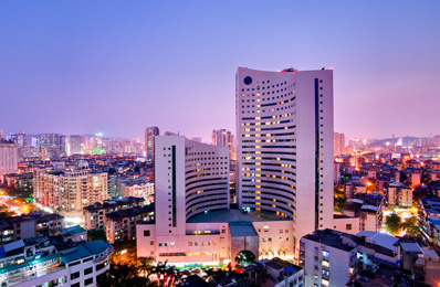
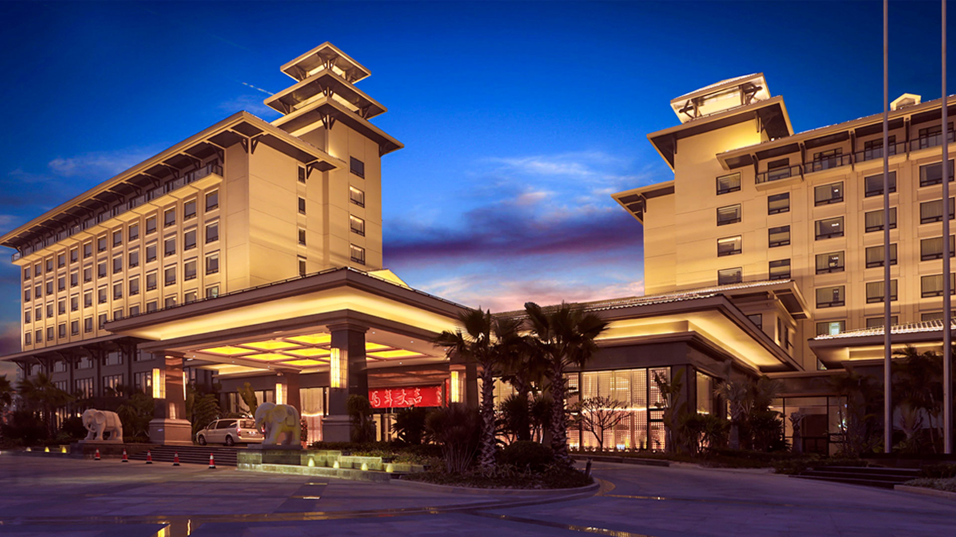
厦门京闽雷速体育(中国)雷速有限公司官网有限公司拥有完善的酒店连锁经营体系
查看详情


1996年
公司成立
01
08
习近平在学习贯彻党的二十大精神研讨班开班式上发表重要讲话习近平,在中央党校建校90周年庆祝大会暨2023年春季学期开学典礼上的讲话。
23
02
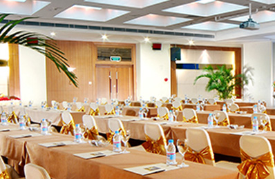
守护年味的服务“坚守者万家灯火时,放弃了个人休息时间以及和小家团聚的时光,让客人们的团聚更加温暖圆满,留守岗位的员工们以饱满的热情接待着来吃团圆饭的宾客们。
09
02
为进一步加强消防安全工作,切实提高全体员工的消防安全意识、防火防灾及突发事件应急处理能力,雷速体育(中国)雷速有限公司官网各下属单位于2024年2月4日、6日及7日分别举行了消防安全演练、安全培训及安全检查等系列活动。
05
02
善谋发展在龙年新春即将到来之际我们用一场年度先进个人;团队颁奖典礼致敬京闽人奋斗进取的2023领导致辞雷速体育(中国)雷速有限公司官网总经理王晖代表公司领导班子,开场舞《好运来》酒店财务部、宾客服务部、餐饮部的小伙伴们将2023年最热门的舞蹈巧妙编排成一支妙趣横生的开门舞。
05
02
福建省能化集团党委书记、董事长徐建平带领集团审计部总经理黄小林、党委宣传部主任谢贤伟莅临雷速体育(中国)雷速有限公司官网公司开展新春慰问,祝福雷速体育(中国)雷速有限公司官网在新的一年里业务越来越好、安全越来越好、管理越来越好。